
Welcome to i-scan
The i-scan imaging project is a collaborative initiative led by PENTAX Medical with Key Opinion Leaders from throughout the endoscopic community. It aims to be a scientific driven platform to gather i-scan experts in a network where clinical cases and i-scan experiences will be shared amongst peers and discussed in frequent webinars.
This website demonstrates the various clinical applications of virtual chromoendoscopy and enhanced imaging with i-scan, in a variety of pathologies in the upper and lower intestine and the bronchial tree. By informing endoscopists of the wide ranging clinical applications and benefits of i-scan imaging, we hope to establish a platform for education, sharing and ultimately providing the highest quality of care to patients.
Sign up for your membership
PENTAX Medical invites our customers to create an account and log in to access to the information about i-scan & OE - Classification Tool, Self-training Lecture, and Webinars
Iaccucci et al
Development and reliability of the new endoscopic virtual chromoendoscopy score: the PICaSSO (Paddington International Virtual ChromoendoScopy ScOre) in ulcerative colitis Journal: GIE. Gastrointestinal Endoscopy. December 2017 Volume 86, Issue 6, Pages 1118–1127.e5
Read moreBowman et al
High Definition Colonoscopy Combined with i-SCAN Imaging Technology Is Superior in the Detection of Adenomas and Advanced Lesions Compared to High Definition Colonoscopy Alone. Diagnostic and Therapeutic Endoscopy
Volume 2015 (2015), Article ID 167406, 5 pages
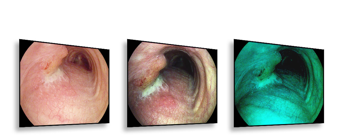
Colonoscopy with one detected polyp in the transverse colon
The video shows 49 year old male with suspected food allergy. During colonoscopy, one polyp in the transverse colon was detected. The histology showed a lymphoid follicle and tubular adenoma with LGIEN.
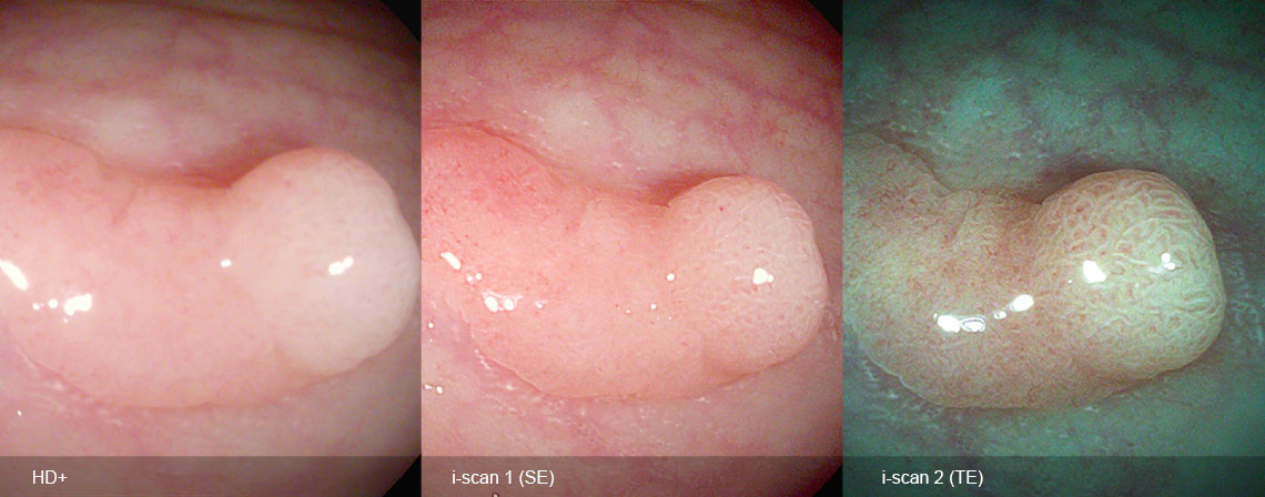
Two aspects in the lesion, the central pit pattern is almost invisible under white light endoscopy and comes out much clearer under i-scan 1 (surface enhancement for detection) and i-scan 2 (tone enhancement for characterization).
Courtesy of Prof. Timo Rath from Erlangen University Hospital, Germany

Colonoscopy with two detected polyps
The video shows a colonoscopy of a 31 year old male with suspected food allergy. During the colonoscopy two polyps were found:
- one tubular adenoma with LGIEN in the descending colon
- one tubular adenoma with LGIEN in the sigmoid colon
The lesion can be easily missed with white light endoscopy (WLE), also in the setting of a very young patient.
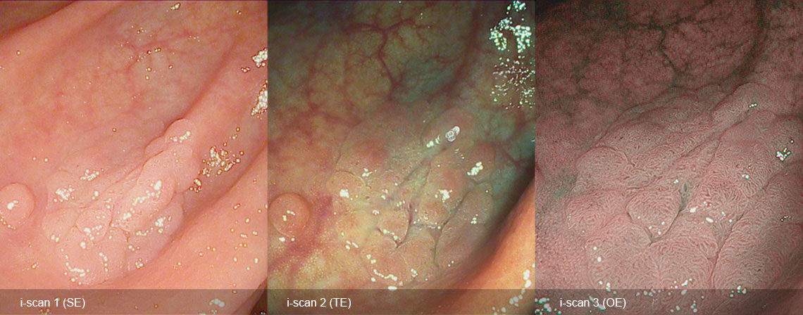
By using i-scan 1, a better demarcation is possible. The vascular pattern are almost invisible unless i-scan OE is used.
Courtesy of Prof. Timo Rath from Erlangen University Hospital, Germany


About i-scan & i-scan OE
A leap forward for in vivo histology with i-scan and i-scan OE, a unique combination of digital and optical enhancements.

i-scan for enhanced detection and characterization
i-scan technology is a digital, image enhanced endoscopy (IEE) technology by PENTAX Medical. As a virtual chromo-endoscopy tool, i-scan provides an enhanced view of the mucosal structures and vascular patterns, supporting early detection, demarcation and characterization.
i-scan OE for vessel characterization
PENTAX Medical has recently developed the i-scan OE (Optical Enhancement), an optical filter that produces bandwidth-limiting light. When combined with image enhancement processing technology it clearly displays the surface structures of blood vessels, glandular ducts and mucosal membranes in higher contrasts than white light.

Optical Imaging (i-scan OE), the newest imaging technology for driving endoscopic therapy in the lower GI tract
Lisbon, May 10th 2016
Courtesy of Prof. Helmut Neumann, University Hospital Erlangen, Germany

In vivo diagnosis of upper GI neoplasia with Optical Imaging (i-scan OE)
Lisbon, May 10th 2016
Courtesy of Dr. Rehan Haidry, University College Hospital London, UK

Brief history of i-scan…and i-scan OE
Lisbon, May 10th 2016
Courtesy of Prof. Dr. Ralf Kiesslich, Wiesbaden, Germany

Imaging Guided Endoscopic Therapy for Early Oesophageal Neoplasia
PENTAX Medical’s UEGW Expert dinner, Camp Nou, Barcelona
Courtesy of Dr. Rehan Haidry, University College Hospital London, UK

Prof. Raf Bisschops
University Hospitals Leuven, Belgium

Professor Helmut Neumann
UK Erlangen, Germany