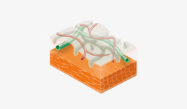
i-scan OE
In addition to the functions that i-scan has offered thus far, namely, Surface Enhancement (SE) or i-scan 1, Tone Enhancement (TE) or i-scan 2 and Contrast Enhancement (CE) or i-scan 3, PENTAX Medical has recently developed an Optical Enhancement (OE) function (hereafter the “i-scan OE”) with the aim of further improving visibility of the blood vessels, ducts of the glands and surface structures. The i-scan OE function incorporates a new technology of combining bandwidth-limiting light with digital image processing. PENTAX Medical’s original optical enhancement (i-scan OE) filters that produce bandwidth-limiting light, combined with image enhancement processing technology, enable the display of the surface structures of the blood vessels, glandular ducts and mucosal membranes in higher contrasts than white light. This combination has the potential to help physicians improve characterization and increase detection and diagnosis of gastrointestinal lesions even more precisely.
The OE filter supports the screening and detailed examination of lesions by using two kinds of modes. Mode 1 provides sufficient light and highlights blood vessels and Mode 2 enhances the blood vessels and the mucosa in a natural color potentially supporting screening.

Gastric cancer
This video sequence demonstrates to observe characteristic of surface pattern with i-scan SE then switch to i-scan OE to observe vessel pattern. i-scan OE brought up better assessment of vessel as compared with chromo endoscopy. Following pathological result concluded mucosal cancer 8mm in size.
Courtesy of Prof.Tomoki Michida, Teikyo University, Japan

Gastric cancer
This video sequence demonstrates to observe characteristic of surface pattern with i-scan SE then switch to i-scan OE to observe vessel pattern and demarcation line of lesion without help of magnified scopes. Following pathological result concluded gastric cancer invaded slightly the submucosa.
Courtesy of Prof.Tomoki Michida, Teikyo University, Japan


