i-scan
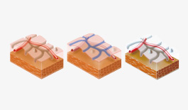
i-scan technology is a digital, real time processing and newly developed image enhanced endoscopy (IEE) technology by PENTAX Medical. As a virtual chromoendoscopy tool, i-scan provides the physician with an enhanced view of the mucosal structures and vascular patterns, supporting early detection, demarcation and characterisation.
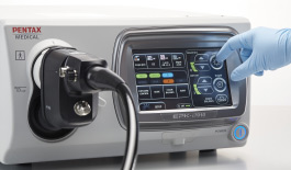
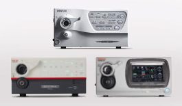
The system is easy to use, quickly engages with the touch of a button, and offers rapid imaging results. It is also possible to display a normal white light image and an i-scan image simultaneously side by side with PENTAX’s unique Twin-mode function provided by its latest HD+ processor, the EPK-i7000. Switching between white light image and the three i-scan settings is quick and can be initialised on a real-time basis.
In GI, HD+ together with i-scan, has been proven to increase adenoma detection rate, meeting the quality measurement standards. During a bronchoscopy, HD+ with i-scan may result in better detection of subtle vascular abnormalities in the airways.


Intramucosal cancer in Barrett`s Oesophagus (Zoom IMC)
This short video shows the exciting new magniview gastroscope in use. This is a patient with a previous Barrett’s Cancer that was resected. At first inspection there appears to be residual flat Barrett’s oeosphagus. However a combination of i-scan and acetic acid shows an area at the right wall with featureless mucosa and abnormal irregular micro-vasculature with zoom endoscopy. One can appreciate the normal adjacent villous mucosa next to the featureless area of intramucosal cancer that was resected by EMR.
Courtesy of Dr. Rehan Haidry, UCLH, UK

Ulcerative colitis
In ulcerative colitis i-scan clearly shows inflammatory changes of the mucosa. We also use it in conjunction with chromoendoscopy for the surveillance of patients with longstanding chronic inflammatory bowel disease to increase detection of flat lesions.
Courtesy of Michael Häfner, St. Elisabeth Krakenhaus, Austria

Crohn`s disease duodenum
Crohn’s disease of the upper GI tract is rare and lesions can easily be missed as they are small. i-scan highlights inflammatory changes of the duodenal mucosa and enhances contrast thus making detection of small aphthoid lesions easier.
Courtesy of Dr. Michael Häfner, St. Elisabeth Krakenhaus, Austria