i-scan OE GI recommended settings
The following settings apply only to the users of the EPK-i7010, as this is the only processor model, which has the i-scan OE (Optical Enhancement) function available.
| Profile i-scan 1 | Profile i-scan 2 | Profile i-scan 3 | |
|---|---|---|---|
| i-scan SE | i-scan TE | i-scan OE 1 | |
| Brightness | 0 | +1 | +3 |
| Ave/Peak | Ave | Ave | Ave |
| Blue | 0 | 0 | 0 |
| Red | 0 | 0 | 0 |
| Enhancement | Low / +2 | Low / +2 | Low / +4 |
| SE | +5 | +4 | NA |
| CE | off | off | NA |
| TE | off | c | NA |
| OE | off | off | Mode 1 |
| Supports detection | Supports pattern characterization and demarcation | Supports vessel characterization |
i-scan 1, uses only SE to refine imaging of subtle surface abnormalities, without altering the brightness of the endoscopic picture.
i-scan 2, combines SE and TE,enhancing minute mucosal changes and vessel structures
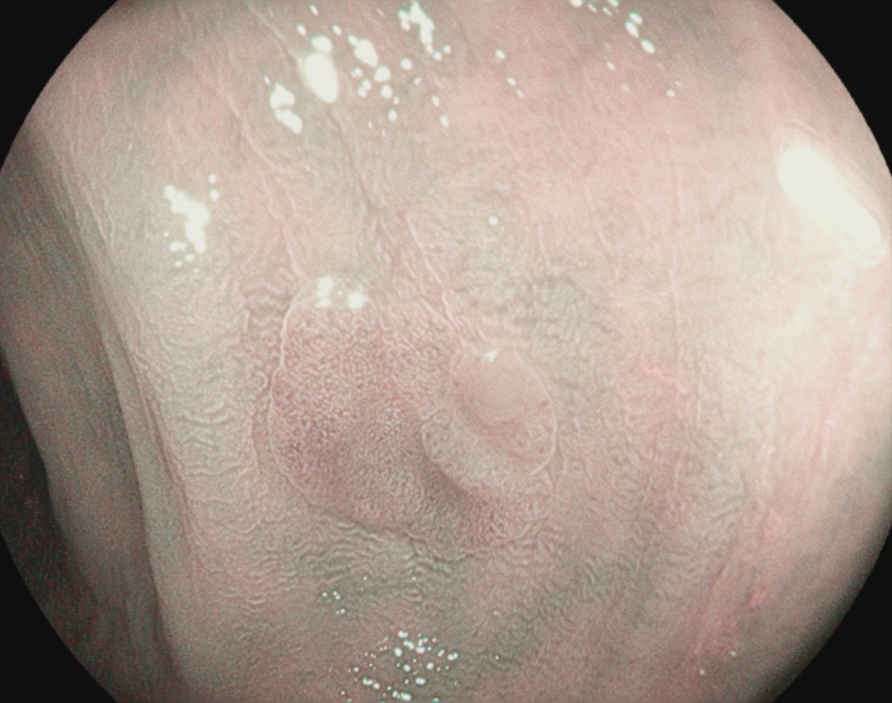
i-scan 3 or i-scan OE 1, optically enhances the mucosal and vascular pattern, adding more information and confidence to the diagonastic
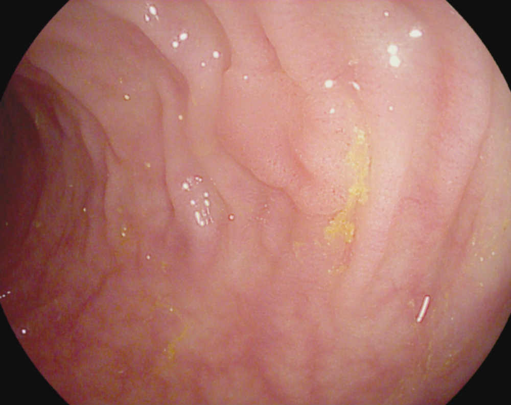
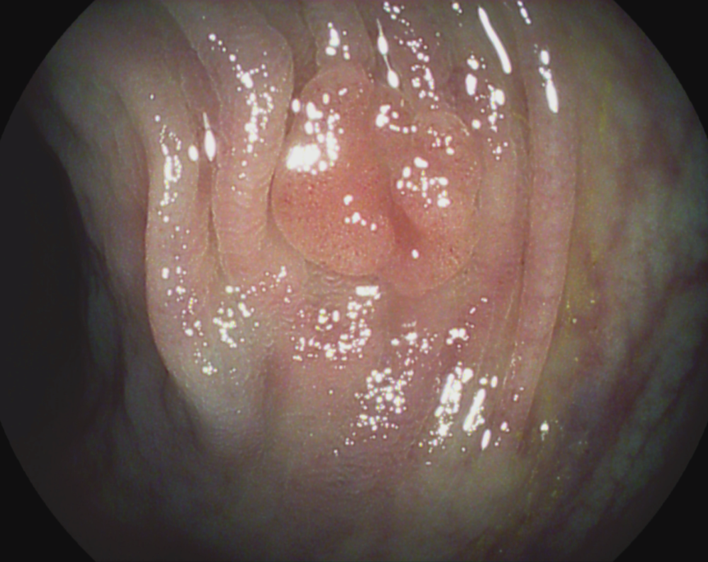
Tubular adenoma with low-grade dysplasia


A subtle change of the epithelial mucosal surface is observed.
The borders of the lesion are clearly delineated from the surrounding mucosa. Combined inspection of the epithelial surface pit-pattern and vascular pit-pattern shows a non-invasive neoplasm. En-bloc EMR was performed


