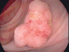
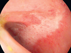
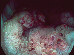
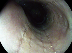
i-scan GI recommended settings
Settings EPK-i5000
| Profile i-scan 1 | Profile i-scan 2 | Profile i-scan 3 | |
|---|---|---|---|
| i-scan SE | i-scan TE | i-scan TE with CE | |
| Brightness | 0 | +1 | +1 |
| Ave/Peak | Ave | Ave | Ave |
| Blue | 0 | 0 | 0 |
| Red | 0 | 0 | 0 |
| Enhancement | Low / +2 | Low / +2 | Low / +2 |
| SE | +5 | +4 | +5 |
| CE | off | off | +2 |
| TE | off | c | c |
| Supports detection | Supports characterisation | Supports demarcation |
Based on current consensus, these are the recommended settings for the three i-scan modes:
(i) i-scan 1 for detection of lesions.
(ii) i-scan 2 for characterisation of lesions.
(iii) i-scan 3 for demarcation of lesions.
i-scan 1, uses only SE to refine imaging of subtle surface abnormalities, without altering the brightness of the endoscopic picture.
i-scan 2, combines SE and TE, enhancing minute mucosal changes and vessel structures.
i-scan 3, in addition to SE and TE, introduces CE to the endoscopic image, digitally adding blue colour to darker edges within the endoscopic image.
Settings EPK-3000 DEFINA
| Profile i-scan 1 | Profile i-scan 2 | Profile i-scan 3 | |
|---|---|---|---|
| i-scan SE | i-scan SE+ TE | i-scan SE + TE | |
| Brightness | 0 | 0 | 0 |
| Ave/Peak | Ave | Ave | Ave |
| Blue | 0 | 0 | 0 |
| Red | 0 | 0 | 0 |
| Enhancement | +5 | +5 | +5 |
| SE | +4 | +4 | +4 |
| CE | off | off | off |
| TE | off | c | g |
| Supports detection | Supports characterization | Supports demarcation |
Based on current consensus, these are the recommended settings for the three i-scan modes:
(i) i-scan 1 for detection of lesions.
(ii) i-scan 2 for characterisation of lesions.
(iii) i-scan 3 for demarcation of lesions.
i-scan 1, uses only SE to refine imaging of subtle surface abnormalities, without altering the brightness of the endoscopic picture.
i-scan 2, combines SE and TE, enhancing minute mucosal changes and vessel structures.
i-scan 3, in addition to SE and TE, introduces CE to the endoscopic image, digitally adding blue colour to darker edges within the endoscopic image.

HD+
- Supports fast orientation and detection
- Significant improvement in the visibility and evaluation of minute lesions
- Mucosal enhancement potentially supports the detection of flat lesions

Detection with i-scan 1 (SE)
- i-scan SE retains the natural color tones
- Accentuation of tissue structures at the touch of a button
- Mucosal enhancement potentially supports the detection of flat lesions

Characterisation with i-scan 2 (TE)
- Specific imaging technology for further assistance in endoscopic procedures
- Allows more accentuated display of mucosal structures which may support lesion characterisation
- Virtual chromoendoscopy may help to improve endoscopic diagnosis

Demarcation with i-scan 3 (TE+CE)
- The depressed area is perceived by blue colour
- Increases visibility of surface structure
- Enhances minute glandular structure change by colour
i-scan in the clinical pathway


