i-scan technology
i-scan consists of three different image algorithms: surface enhancement (SE); tone enhancement (TE); and contrast enhancement (CE).
The three modes (SE, TE and CE) are arranged in series, therefore, it is possible to apply two or more of these modes at one time. Switching the levels or modes of enhancement can be performed on a real-time basis, without any time lag, by pushing a scope button, enabling efficient endoscopic observation.
Surface enhancement (SE) | Contrast enhancement (CE) | Tone enhancement (TE)
Surface Enhancement improves light-dark contrast by obtaining luminance intensity data for each pixel and applying an algorithm that allows detailed observation of the mucosal surface structure and lesion borders in a natural colour tone.
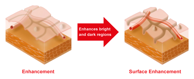
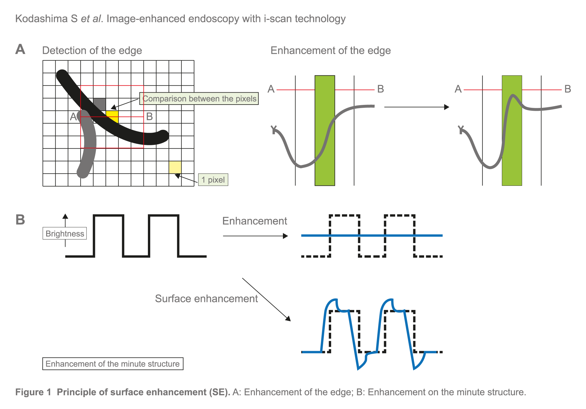
With SE, the difference in luminance intensity between the pixels concerned and the surrounding pixels is analysed, and the edge components are enhanced (Figure 1A). With ordinary enhancement, minor changes in structure are perceived as noise, and the area that shows such changes is smoothed out. With SE, adjustment of the noise erasure function allows more evident enhancement of the edges, which corresponds to minor changes in structure (Figure 1B).
Focal hyperplasia of the gastric mucosa
WL Image
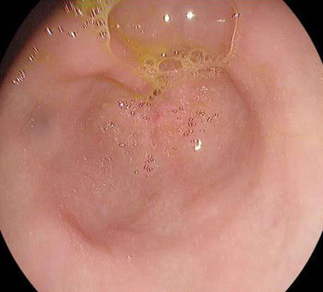
SE Image
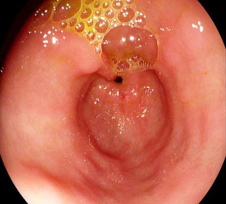
Courtesy of Dr Irina Khimina, KDC №6, Russia
Hypervascular mass in the right upper lobe
WL Image
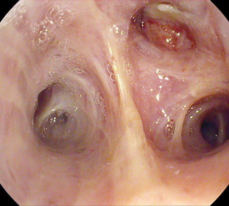
SE Image
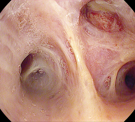
Courtesy of Dr. Javier Cosano, Hospital Reina Sofía de Córdoba, Spain
Contrast enhancement (CE) (Used only in GI i-scan settings) | Surface enhancement (SE) | Tone enhancement (TE)
CE digitally adds blue colour to relatively dark areas within the endoscopic image by obtaining luminance intensity data for each pixel. It then applies an algorithm which allows detailed observation of subtle irregularities around the surface and a detailed inspection of the mucosal vascular pattern morphology.
Even in the case of flat mucosa, the minute glandular structure can be enhanced because the colour is changed, which reflects very small depressions at the gland duct opening. Processing of images with CE does not cause a change in image brightness or a marked change in the colour of the images, it causes only slight bluish-white staining of depressed areas.
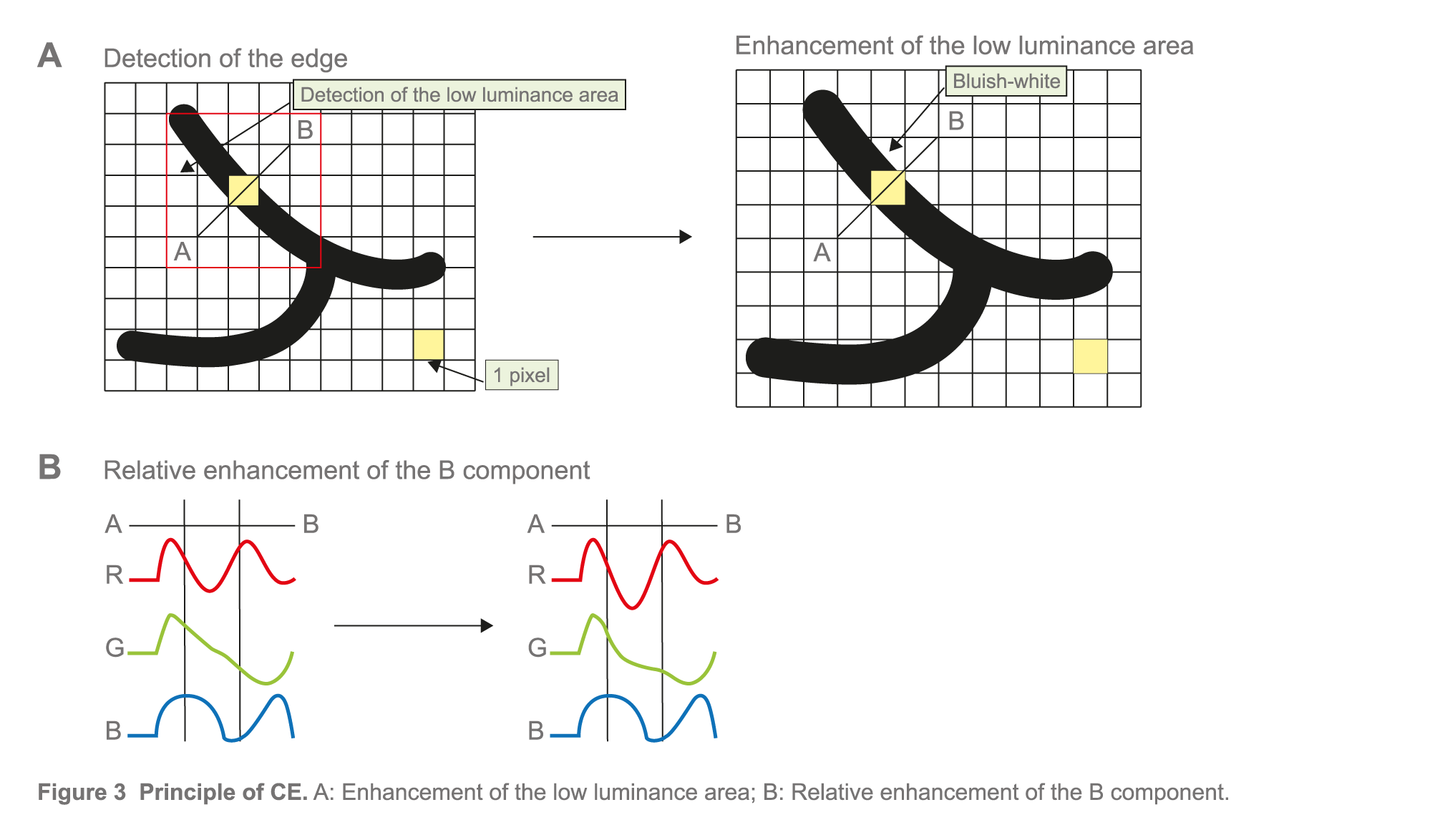
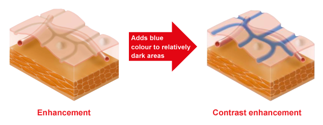
Both SE and CE functions can be switched between three enhancement levels - low, medium and high – and work in real-time without impairing the original colour of the organ. Therefore both functions are suitable for screening endoscopy to detect gastrointestinal tumours at an early stage1.
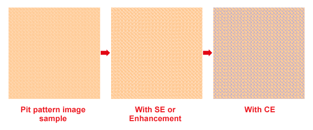
i-scan images are as bright as conventional white light images, allowing much larger areas to be observed in a distant view compared with NBI. Consequently, i-scan can be more useful for performing screening endoscopy and diagnosing the extent of a lesion, particularly that of a large superficial lesion in the stomach1.
i-scan can be used simply and instantaneously by pushing a button, providing an easy method for screening or detailed inspection, and may reduce both time and costs when compared to traditional chromoendoscopy1.
Tone enhancement (TE) | Surface enhancement (SE) | Contrast enhancement (CE)
TE dissects and analyses the individual red-green-blue (RGB) components of a normal endoscopic image in real-time, alters the colour frequencies of each component, and recombines the components to a single, new colour image without visible delay for the examiner (Figure below). Thereby, TE enhances mucosal structures, vascular patterns and subtle changes in colour.
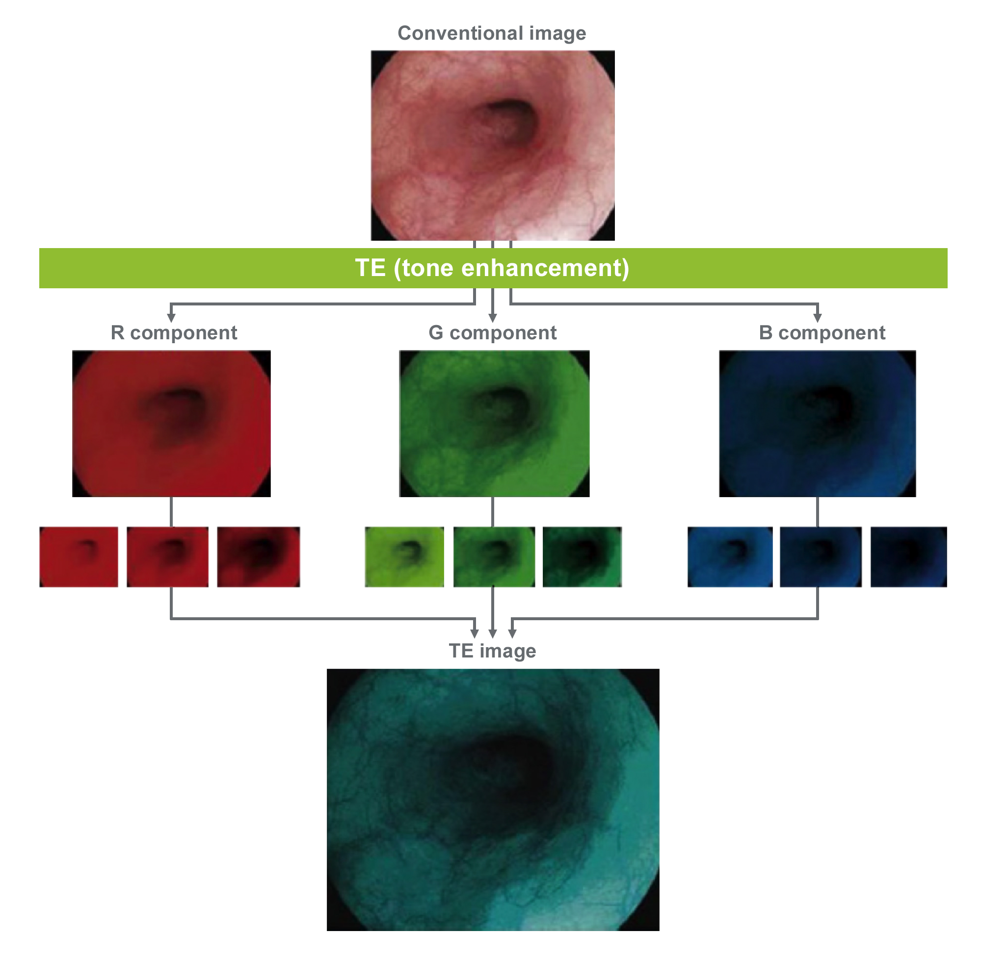
TE works in real time and can be used in 5 modes: TE [p] for pit pattern, TE [v] for vascular pattern, TE [b] for Barrett’s, TE [e] for oesophagus, TE [g] for stomach and TE [c] for colon.
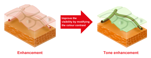
Focal hyperplasia of the gastric mucosa
WL Image

TE Image
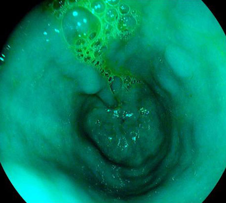
Courtesy of Dr Irina Khimina, KDC №6, Russia
Hypervascular mass in the right upper lobe
WL Image

TE Image
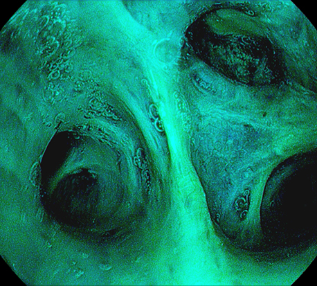
Courtesy of Dr. Javier Cosano, Hospital Reina Sofía de Córdoba, Spain
1Kodashima et al: Novel image-enhanced endoscopy with i-scan technology. World J Gastroenterol 2010
Downloads
A unique combination of optical and digital enhancements, for improved in vivo diagnosis
A leap forward for in vivo histology with i-scan and i-scan OE, a unique combination of digital and optical enhancements
i-scan, technology with proven results
A novel endoscopic classification system using i-scan improves dysplasia detection in Barrett’s oesophagus
PENTAX Medical i-scan Mini-Atlas for Gastroenterology
This i-scan Mini-Atlas will give you a comprehensive overview about the imaging options of i-scan based on selected clinical cases of the upper and lower GI-tract.
Gaining a complete picture of the GI tract through new image enhancement technology
i-scan real-time image processing technology has proven to be a clinically-effective digital image enhanced endoscopy (IEE) technique, using virtual chromoendoscopy to provide superior visualization of vascular and tissue architecture on the mucosal surface. Now a new optical enhancement option using band-limited light has been added to the i-scan menu.
